Resonant X-Ray Diffraction (RXD)
X-ray diffraction (XRD) is one of the essential tools for studying crystal structures, using interference between incident and scattered x-ray lights. Resonant x-ray diffraction (RXD) combines XRD and x-ray absorption spectroscopy, which can be achieved by tuning the incident photon energy to atomic x-ray absorption edges. This tuning process, coined ‘resonant process’, greatly enhances the scattering cross-sections and allows careful investigation of various ordering phenomena hardly observable with conventional XRD. Furthermore, the element specificity (obtained by selecting an absorption edge) and capability for small crystals (~100 um) make RXD distinguished among other diffraction techniques including magnetic neutron diffraction.
In strongly correlated electronic systems, a myriad of exotic states are manifested by ordering phenomena; for example, loss of rotational and translational symmetry can appear as nematic and charge density wave (CDW) orders, respectively. These ordering phenomena can be best explored by RXD, and we mainly focus on transition metal oxide compounds, one of the largest family of the strongly correlated electronic systems.
We’re in collaboration with RXD beamlines all around the world, including sector 4 and 6 at Advanced Photon Source (APS) in USA and P09 at Deutsches Elektronen-Synchrotron (DESY) in Germany.
Resonant Inelastic X-Ray Scattering (RIXS)
Resonant inelastic x-ray scattering (RIXS) is a photon-in photon-out process that can measure bosonic excitations in a solid, resolving their momenta and energies. They include various elementary excitations, such as plasmons, charge-transfer, charge-density waves, crystal-field excitations, magnetic excitations, etc. Particularly, ever since single-magnon excitations are resolved by RIXS, as demonstrated by Ament and Braicovich [1,2], RIXS technique has achieved remarkable progress especially in its energy resolution (<100 meV) and count rate (>103 sec-1).
Moreover, RIXS can choose specific excitations to be measured, amplifying their cross-section through resonating the atomic edges. For example, magnons hardly visible with conventional x-ray tools can dominate the spectra when the light resonates the magnetic ions (see the RIXS data below).
Our group uses state-of-the-art hard x-ray RIXS beamlines around the world (including 27ID in APS and the 20ID in ESRF) to study magnetic properties of the iridium oxides using Ir L3-edge (11.215 keV) enjoying their great energy-resolution (~25 meV), x-ray intensities and beam focusing. After their planned upgrades, x-ray intensities and beam focusing will be further enhanced, providing more chances to study samples under extreme environments such as pressure and magnetic fields. Furthermore, we are also constructing our own hard x-ray RIXS beamline at 1C in PLS-II which focuses on the Ir L3-edge, aiming one of the best hard x-ray beamlines in the world.

[1] L. J. P. Ament et al., PRL 103, 117003 (2009).
[2] L. Braicovich et al., PRL 104, 077002 (2010).
Thermal Diffuse Scattering
to be updated
Angle-resolved photoemission spectroscopy (ARPES)
Angle-resolved photoemission spectroscopy (ARPES) directly probes the electronic band structure of matter based on the principle of the photoelectric effect. When the incident photon expels electrons in solids, the electrons are captured by the spectrometer which analyzes their dispersion relation. Since the photo-excited electrons are dressed by various interactions in the solid, the dispersion carries fruitful information about the spin, orbital, and topological properties of the matter. Therefore, ARPES becomes one of the most potent and essential techniques in modern solid-state physics. ARPES is widely employed to study various exotic materials including strongly-correlated system, Dirac materials, and two-dimensional materials.

Raman spectroscopy
Raman spectroscopy is an inelastic light scattering procedure, measuring energy difference between the incident and scattered lights. It reveals various excitations as sharp features in the energy spectrum, whose positions indicate energy cost (gain) for their creation (annihilation). Raman spectroscopy usually uses visible-light laser, and this high-flux laser discloses a plethora of excitations in a solid, such as optical phonons, magnons, crystal field excitations, inter-band excitations, and their pair-creations, with an exceptional resolution ( ~ 1 cm-1) at the Brillouin zone center (q = 0). Thus, it is an indispensable tool for studying condensed matter systems, and nowadays, commercialized instruments come into wide use.
Our home-built Raman setup has several advantages beyond the commercial instruments: high resolution, capability of polarization analysis, and resonance studies. First, our system uses a long-focal-length (75 cm) spectrometer and Bragg-grating notch filters to finely resolve (~ 0.1 meV resolution) low-energy (down to ~ 1 meV) excitations. Second, it controls polarization of the incident and scattered light, realizing various polarization configurations in a back-scattering geometry. The polarization dependencies provide extensive information about symmetry of the excitations. Third, our system is equipped with different colored lasers (488 nm, 532 nm, and 633 nm), which can investigate resonant properties of the excitations. The Raman signals are significantly amplified when the incident photon energy approaches their intermediate states that are closely related to the underlying dynamics. Our setup also includes closed-cycle LHe cryostat cooling down the crystals to 4.2 K.

Rotational Anisotropy Second Harmonic Generation (RA-SHG)
Rotational anisotropy second harmonic generation (RA-SHG) is highly sensitive to electronic and structural symmetry breaking in a crystal. When optical pulses are incident on the crystal surface, it collects second-harmonic-generation (SHG) signals of the twice photon energy using a 2D CCD detector. Each CCD pixel measures SHG signal at different incidence orientation (which also has two-dimensional degrees of freedom θ, φ). Therefore, when the scattering plane rotates about the surface normal at a fixed θ, the SHG signal circles on the CCD, resulting in a circular pattern that visualizes the crystalline symmetry. The crystalline symmetry can be carefully examined by the best fits to the circular patterns, and usually, four different patterns are obtained by aligning the incident / scattered polarizations to be parallel / perpendicular to the scattering plane.

Crystal Synthesis
Preparation of single crystalline sample is the most fundamental step in the studies of condensed matter physics, yet it entails many difficulties that requires multidisciplinary approaches for the solutions. Our group intends to examine the growths of transition-metal oxide crystals in multiple facets by implementing techniques ranging from solution-based methods such as the flux methods, to vapor-based methods such as chemical-vapor transport (CVT).
Our synthesis facility includes a number of furnaces capable of reaching temperatures up to 1500 ℃ in controlled atmospheres; and auxiliary equipment for handling air-sensitive materials. Characterizations are usually done through x-ray diffraction (XRD) and the energy-dispersive x-ray spectroscopy (EDXS). Along with the applications and optimizations of the existing techniques for our materials of interest, we are also actively developing new synthesis techniques catered to various phases of transition-metal oxides. The current objective for the synthesis sector of our research is focused towards the controlled growth and doping of iridates (i.e. iridium-based oxides).

Aerosol-assisted Chemical Vapor Deposition
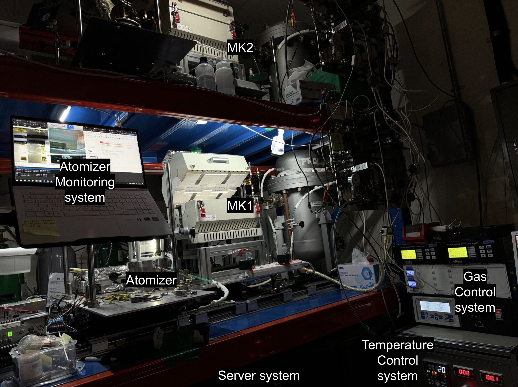
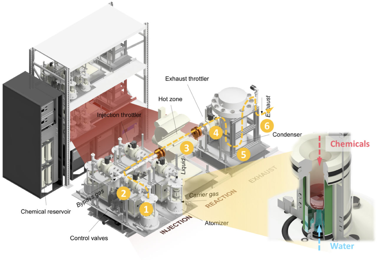
Our lab has developed and refined a unique aerosol-assisted chemical vapor deposition (AACVD) system specifically designed for synthesizing high-quality single crystals of complex transition metal oxides, including materials like Sr₂IrO₄. The AACVD technique utilizes a fine aerosol mist containing precursor materials, which is carried by an inert gas or diluted oxygen into the reactor chamber. There, it decomposes at elevated temperatures, forming a powder phase, thin film, or single crystal directly on the substrate. This setup is particularly advantageous for controlling stoichiometry, allowing precise adjustments in growth parameters, and resulting in reproducible, high-quality crystals with excellent phase purity and well-defined morphology.
Unlike traditional chemical vapor deposition, AACVD introduces material precursors in a liquid phase, which expands the range of compounds that can be synthesized with greater uniformity and scalability. Our system stands out globally due to its unique capability of achieving homogeneous deposition across larger areas while maintaining atomic precision. This innovation enables in-depth studies of magnetism and complex electronic behaviors in transition metal oxides and other quantum materials.

